According to the American Board of Surgery, the specialty of general surgery is defined as: General surgery is a discipline that requires knowledge of and familiarity with a broad spectrum of diseases that may require surgical treatment. By necessity, the breadth and depth of this knowledge will vary by disease category. In most areas, your Ocala surgeon will be expected to be competent in diagnosing and treating the full spectrum of disease. However, there are some types of disease in which comprehensive knowledge and experience is not generally gained in the course of a standard surgical residency. In these areas, the surgeon will be able to recognize and treat a select group of conditions within a disease category.
The team at Surgical Specialists of Ocala have successfully pursued training in the following areas of study to serve their patients to the highest level possible in Ocala, The Villages, and the surrounding Central Florida areas:

The appendix plays an important role in of the function of the human anatomy. It produces a protein called immunoglobulins that destroy bacteria and help fight infection. While the appendix is important, it’s not essential. Other organs in the body compensate when an appendix must be removed. Just like a blind person’s other senses increase to compensate for no vision. The human anatomy is remarkable. People who have had appendectomies do not have an increased risk toward infection.
Appendicitis is very common, experienced by one of every 2,000 people per year. It occurs when the appendix becomes inflamed. The inflammation causes discomfort. When this occurs, prompt removal of the appendix is necessary. If the inflammation is allowed to continue, the appendix could burst or perforate, spilling infectious materials into the abdominal cavity.
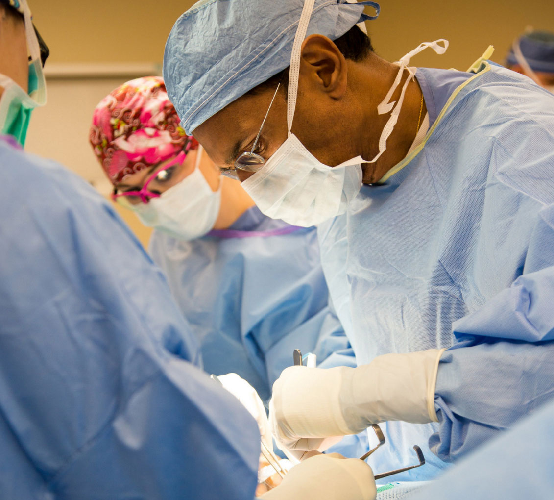
As with any procedure they perform, Surgical Specialists of Ocala choose to perform appendectomies with the least invasive technique possible. A laparoscopic procedure is the first choice due to the following advantages:
Although laparoscopic appendectomy has many benefits, it may not be appropriate for some patients. Early, non-ruptured appendicitis usually can be removed laparoscopically. Laparoscopic appendectomy is more difficult to perform if there is advanced infection or the appendix has ruptured. A traditional, open procedure using a larger incision may be required to safely remove the infected appendix in these patients.
Surgical Specialists of Ocala work closely with other specialists to determine the timing and procedures needed to offer patients the best chance for victory in the battle against breast cancer.
When you and/or your doctor find a lump during a breast examination or a suspicious area found on a mammogram, ultrasound, or magnetic resonance imaging (MRI), your doctor will most likely recommend a biopsy. This removal of a small portion of the mass is reviewed under a microscope to determine whether or not cancerous cells exist.
There are several ways Surgical Specialists of Ocala perform breast biopsies:
If needed, your doctor may use ultrasound or MRI to guide the biopsy needle. Or your doctor may use a computer to locate the exact spot for the biopsy sample from mammograms that have been taken from two angles (stereotactic needle biopsy). A fine wire, clip, or marker also may be used to mark the site.
Lumpectomy is surgery to remove cancer or other abnormal tissue from your breast. Lumpectomy is also called breast-conserving or breast-sparing surgery because — unlike a mastectomy — only a portion of the breast is removed. Doctors may also refer to lumpectomy as a wide local excision. During lumpectomy, a small amount of normal tissue around the lump (clean margins) also is taken to help ensure that all the cancer or other abnormal tissue is removed.
Lumpectomy helps confirm a diagnosis of cancer or rule it out. Lumpectomy is also a first treatment option for some women with early-stage breast cancer. In cases where cancer is found, lumpectomy usually is followed by radiation therapy to reduce the chances of cancer returning.
Mastectomy is surgery to remove all breast tissue from a breast as a way to treat or prevent breast cancer.
For those with early-stage breast cancer, mastectomy may be one treatment option. Breast-conserving surgery (lumpectomy), in which only the tumor is removed from the breast, may be another option. Deciding between mastectomy and lumpectomy can be difficult. Both procedures are equally effective. But lumpectomy isn’t an option for everyone with breast cancer, and others prefer to undergo a mastectomy.
Newer mastectomy techniques can preserve breast skin and allow for a more natural breast appearance following the procedure. Surgery to restore shape to your breast — called breast reconstruction — may be done at the same time as your mastectomy or during a second operation at a later date.
The colon is the large intestine; it is the lower part of your digestive tract. The intestine is a long, tubular organ consisting of the small intestine, the colon (large intestine) and the rectum, which is the last part of the colon. After food is swallowed, it begins to be digested in the stomach and then empties into the small intestine, where the nutritional part of the food is absorbed. The remaining waste moves through the colon to the rectum and is expelled from the body. The colon and rectum absorb water and hold the waste until you are ready to expel it.
Damage or disease in the colon requires a surgical procedure to remove affected section to prevent further spread of disease and discomfort. Surgical Specialists of Ocala prefer to preform colon resections laparoscopically. It is the least invasive procedure with a quicker recovery. Not all patients are candidates for laporascopic colon resection and an “open” procedure is required. Surgical Specialists of Ocala will always defer to the safest and most effective option for each patient.
The gallbladder is a pear-shaped organ that rests beneath the right side of the liver. Its main purpose is to collect and concentrate a digestive liquid (bile) produced by the liver. Bile is released from the gallbladder after eating, aiding digestion. Bile travels through narrow tubular channels (bile ducts) into the small intestine. Removal of the gallbladder is not associated with any impairment of digestion in most people. Gallbladder removal is one of the most commonly performed surgical procedures in the United States. Today, gallbladder surgery is performed laparoscopically. The medical name for this procedure is Laparoscopic Cholecystectomy.
Gallbladder problems are usually caused by the presence of gallstones: small hard masses consisting primarily of cholesterol and bile salts that form in the gallbladder or in the bile duct. It is uncertain why some people form gallstones. There are no known means to prevent gallstones. These stones may block the flow of bile out of the gallbladder, causing it to swell and resulting in sharp abdominal pain, vomiting, indigestion and, occasionally, fever. If the gallstone blocks the common bile duct, jaundice (a yellowing of the skin) can occur.
When gallbladder removal is necessary, Surgical Specialists of Ocala prefer to preform it laparoscopically. It is the least invasive procedure with a quicker recovery. Not all patients are candidates for laparoscopic gallbladder surgery and an “open” procedure is required. Our Ocala surgeons will always defer to the safest and most effective option for each patient.
Gastrointestinal diseases refer to diseases involving the gastrointestinal tract, namely the esophagus, stomach, small intestine, large intestine and rectum, and the accessory organs of digestions, the liver, gallbladder, and pancreas.
The lining of the esophagus (food pipe) is a soft, moist tissue called mucosa. When stomach acids or in some cases stomach content, flows back into the esophagus it irritates the soft lining and causes a chronic digestive disease called Gastroesophageal Reflux Disease (GERD).
Occasional acid reflux and heartburn are common digestive conditions that are not usually reason for concern. However, when these signs and symptoms occur at least twice each week, interfere with your daily life, or when your doctor can see damage to your esophagus, you may be diagnosed with GERD.
Most people can manage the discomfort of GERD with lifestyle changes and over-the-counter medications. But some people with GERD may need stronger medications, or even surgery, to reduce symptoms.If your doctor recommends surgery, Ocala Surgical Specialists are committed to taking care of your condition in the least invasive way.
Where dietary changes and medications have not improved GERD, Surgical Specialists of Ocala may perform a Laparoscopic anti-reflux surgery (commonly referred to as Laparoscopic Nissen Fundoplication) involves strengthening the “valve” between the esophagus and the stomach by wrapping the upper portion of the stomach around the lowest portion of the esophagus.
In a laparoscopic procedure, surgeons use small incisions (1/4 to 1/2 inch) to enter the abdomen through cannulas (narrow tube-like instruments). The laparoscope, which is connected to a tiny video camera, is inserted through the small incision, giving the surgeon a magnified view of the patient’s internal organs on a television screen.
The entire operation is performed “inside” after the abdomen is expanded by inflating gas into it.
In some cases, this surgery may require an “open” vs. Laproscopic procedure. Surgical Specialists of Ocala weigh all factors carefully, keeping the patient’s safety and ease of recovery in mind.
A hernia is the protrusion of an organ or part of an organ through the wall of the cavity that normally contains it. A hernia occurs when there is a weakness or tear in the muscle wall as a result of aging, injury, a previous surgical incision, or a condition present at birth. Hernias generally grow larger due to pressure on them, such as a loop of your intestine or fatty tissue pushing into the weak abdominal tissue or tear. The result is pressure on that area causing the tissue or organ to push through and bulge out. Depending on the location and severity of your hernia, as well as your medical history, your doctor will recommend either open or laparoscopic surgery. Within each surgical option are different hernia repair techniques:
Also known as a “sutured repair”, this repair involves sewing the abdominal wall tissues back together.
A patch is attached over the weak area around the hernia in front of the muscles. Over time, your body’s tissue grows naturally into the patch to make a strong repair.
A space-filling plug is placed inside the inguinal hernia to reinforce and support the weak tissue. A patch is then attached over the area.
The skin incision is closed with stitches, staples, surgical tape, or special glue.
The main symptom of an inguinal hernia is a bulge in the groin or scrotum. It often feels like a round lump. The bulge may form over a period of weeks or months. Or it may appear all of a sudden after you have been lifting heavy weights, coughing, bending, straining, or laughing. The hernia may be painful, but some hernias cause a bulge without pain.
An incisional hernia is a type of hernia caused by an incompletely-healed surgical wound. Since median incisions in the abdomen are frequent for abdominal exploratory surgery, ventral incisional hernias are often also classified as ventral hernias due to their location.
An umbilical hernia is a bulge or pouch that forms in the abdomen. This bulge occurs when a section of the lining of your abdominal cavity pushes through the abdominal wall around where the belly button is located. Umbilical hernias are common in children and adults.
A rare and serious complication related to umbilical hernias in adults is called strangulation. Strangulation is when the blood flow to certain parts of the body is cut off because the hernia causes tissue to bulge. Symptoms of strangulation include nausea, vomiting, and severe pain. The area around the umbilical hernia might look blue, as if you have a bruise. If it does look like this, call a doctor or healthcare professional right away.
Ventral hernias are a type of abdominal hernia. They may develop as a defect at birth, resulting from incomplete closure of part of the abdominal wall, or develop where an incision was made during an abdominal surgery, occurring when the incision doesn’t heal properly.
The Vein Center of Ocala uses the latest technology and least invasive procedures possible to repair and optimize the function of your veins. It’s not about vanity, it’s about vitality. What you see on the outside means correction is needed on the inside. The Vein Center of Ocala can help.